Biology
Causal Agent:
Fascioliasis is caused by Fasciola hepatica and less often by F. gigantica, which are flat worms classified as liver flukes (trematodes). Some human cases have been caused by hybrid species. Additional Fasciola species have been found in animals.
Life Cycle:
As shown below, Fasciola parasites develop into adult flukes in the bile ducts of infected mammals, which pass immature Fasciola eggs in their feces. The next part of the life cycle occurs in freshwater. After several weeks, the eggs hatch, producing a parasite form known as the miracidium, which then infects a snail host. Under optimal conditions, the development process in the snail may be completed in 5 to 7 weeks; cercariae are then shed in the water around the snail. The cercariae lose their tails when they encyst as metacercariae (infective larvae) on water plants. In contrast to cercariae, metacercariae have a hard outer cyst wall and can survive for prolonged periods in wet environments.
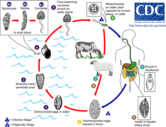
Immature Fasciola eggs are discharged in the biliary ducts and in the stool  . Eggs become embryonated in water
  , eggs release miracidia
  , which invade a suitable snail intermediate host
  , including the genera Galba, Fossaria and Pseudosuccinea. In the snail the parasites undergo several developmental stages (sporocysts
  , rediae
  , and cercariae
  ). Thecercariae are released from the snail
  and encyst as metacercariae on aquatic vegetation or other surfaces. Mammals acquire the infection by eating vegetation containing metacercariae. Humans can become infected by ingesting metacercariae-containing freshwater plants, especially watercress
  . After ingestion, the metacercariae excyst in the duodenum
  and migrate through the intestinal wall, the peritoneal cavity, and the liver parenchyma into the biliary ducts, where they develop into adult flukes
  .
.
In humans, maturation from metacercariae into adult flukes takes approximately 3 to 4 months. The adult flukes (Fasciola hepatica: up to 30 mm by 13 mm; F. gigantica: up to 75 mm) reside in the large biliary ducts of the mammalian host. Fasciola hepatica infect various animal species, mostly herbivores (plant-eating animals).
Disease
Human fascioliasis is usually recognized as an infection of the bile ducts and liver, but infection in other parts of the body can occur.
In the early (acute) phase, symptoms can occur as a result of the parasite's migration from the intestine to and through the liver. Symptoms can include gastrointestinal problems such as nausea, vomiting, and abdominal pain/tenderness. Fever, rash, and difficulty breathing may occur.
During the chronic phase (after the parasite settles in the bile ducts), the clinical manifestations may be similar or more discrete, reflecting inflammation and blockage of bile ducts, which can be intermittent. Inflammation of the liver, gallbladder, and pancreas also can occur.
Diagnosis
The standard way to be sure a person is infected withFasciola is by seeing the parasite. This is usually done by finding Fasciola eggs in stool (fecal) specimens examined under a microscope. More than one specimen may need to be examined to find the parasite. Sometimes eggs are found by examining duodenal contents or bile.
Infected people don't start passing eggs until they have been infected for several months; people don't pass eggs during the acute phase of the infection. Therefore, early on, the infection has to be diagnosed in other ways than by examining stool. Even during the chronic phase of infection, it can be difficult to find eggs in stool specimens from people who have light infections.
Certain types of blood tests can be helpful for diagnosingFasciola infection, including routine blood work and tests that detect antibodies (an immune response) to the parasite.
Treatment
The first step is to make sure the diagnosis is correct. For more information, patients should consult their health care provider. Health care providers may consult with CDC staff about the diagnosis and treatment of fascioliasis.
The drug of choice is triclabendazole. In the United States, this drug is available through CDC, under a special (investigational) protocol. The drug is given by mouth, usually in one or two doses. Most people respond well to the treatment.
Prevention & Control
No vaccine is available to protect people against Fasciola infection.
In some areas of the world where fascioliasis is found (endemic), special control programs are in place or are planned. The types of control measures depend on the setting (such as epidemiologic, ecologic, and cultural factors). Strict control of the growth and sale of watercress and other edible water plants is important.
Individual people can protect themselves by not eating raw watercress and other water plants, especially from endemic grazing areas. As always, travelers to areas with poor sanitation should avoid food and water that might be contaminated (tainted). Vegetables grown in fields that might have been irrigated with polluted water should be thoroughly cooked, as should viscera from potentially infected animals.